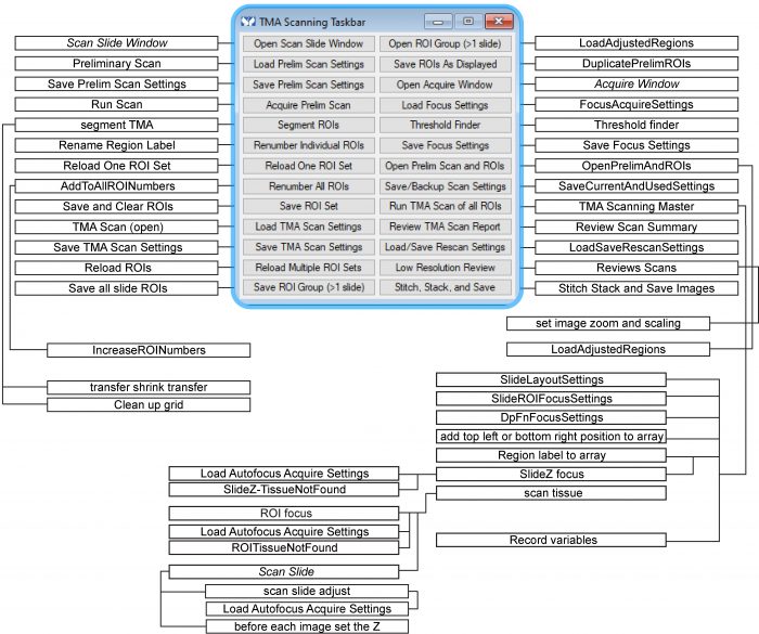
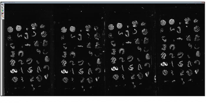
High-throughput, high-content imaging technologies and multiplex slide scanning have become widely used. Advantages of these approaches include the ability to archive digital copies of slides, review slides as teams using virtual microscopy software, and standardize analytical approaches. The cost and hardware and software inflexibility of dedicated slide scanning devices can, however, complicate implementation. Here, we describe a simple method that allows any microscope to be used for slide scanning. The only requirements are that the microscope be equipped with a motorized filter turret or wheels (for multi-channel fluorescence) and a motorized xyz stage. This example uses MetaMorph software, but the same principles can be used with any microscope control software that includes a few standard functions and allows programming of simple command routines, or journals. The series of journals (and their text, as a word document) that implement the method perform key functions, including assistance in defining an unlimited number of regions of interest (ROI) and imaging parameters. Fully automated acquisition is rapid, taking less than 3 hr to image fifty 2.5-mm ROIs in four channels. Following acquisition, images can be easily stitched and displayed using opensource or commercial image-processing and virtual microscope applications. Results using the method described are available within the Atlas of Intestinal Transport.